In this condition, also known as isidiopathic hypertrophic subaortic stenos, there is cardiac hypertrophy, usually involving the interventricular septum, which in a minority of patients can lead to a dynamic left ventricular outflow obstruction. In the patients with obstruction any situation that results in a reduction in left ventricular volume (hypovolemia, Valsalva maneuver) increases the obstruction of the thickened, non-compliant, “restrictive” ventricle. The disease has a genetic basis, at times affecting families, and involves mutations in the coding of b cardiac myosin heavy chains. The majority of patients are asymptomatic but the condition can present, usually in young adults, with syncope, sudden death, or breathlessness. Abnormal cardiac rhythms are poorly tolerated and patients may present with atrial tachyarrhythmias, leading to hypotension, as a result of the loss of the atrial contribution to filling the “restrictive” ventricle.
On physical examination, the pulse is bisferiens or “jerky”, with a brisk arterial upstroke. Blood pressure is normal and JVP is usually normal. There is a prominent, forceful, left ventricular impulse.
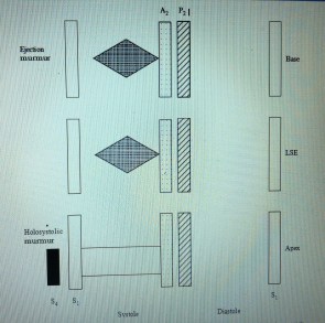
 On auscultation (Figure), S1 is normal and S2 is split normally on inspiration. There is a prominent S4. A mid to late systolic ejection murmur is heard at the left sternal edge and apex. The murmur typically increases with Valsalva maneuver. When mitral regurgitation is present, an apical, holosystolic murmur is heard and may be accompanied by an S3.
The chest and abdominal examinations are normal. There is no peripheral edema. Electrocardiogram findings are left ventricular hypertrophy, ST and T wave abnormalities, and prominent septal Q
| Feature | Obstructive hypertrophic cardiomyopathy | Aortic valve stenosis |
| Pulse | Bisferiens or “jerky”, with a brisk | Slow rising, low volume, |
| ar terial upstroke | and sustained | |
| Murmur | Ejection systolic; increases with | Ejection systolic; |
| Valsalva (which decreases stroke | increases with squatting | |
| volume) with or without mitral | (which increases stroke | |
| holosystolic | volume) |
Distinguishing examination features of obstructive hypertrophic cardiomyopathy and aortic valve stenosis
waves (narrow and deep) caused by hypertrophy of the septal region (a “pseudo-infarction” appearance). The chest x ray film appearance is normal or cardiac enlargement may be present. Certain examination features distinguish obstructive hypertrophic cardiomyopathy (subaortic stenosis) from aortic valve stenosis (Table above).
The clinician needs to be aware that hypertrophic obstructive myopathy can be found in young adults; it may be unmasked by inappropriate hypotension associated with atrial arrhythmias (because of restriction of cardiac filling), and the obstructive variety often has an S4 and an ejection systolic murmur.
Source: Cardiology Core Curriculum A problem-based approach John D Rutherford


