A 6-year-old male developed an upper respiratory tract infection followed by facial edema with dark-colored urine after 2 weeks. Upon examination, his blood pressure was at 120/80 mmHg. There were hyperpigmented lesions noted on the lower extremities were probably due to previous active skin infection. Urinalysis reveals too numerous to count RBCs/hpf, 8-10 WBCs/hpf and 4+ protein. Phase contrast microscopy showed >80% dysmorphic red cells.
QUESTIONS:
1. What is the etiology of AGN in this case?
The most common infectious cause of acute GN is infection by Streptococcus species (i.e. group A, beta-hemolytic). Two types have been described, involving different serotypes:
-
Serotype 12 – Poststreptococcal nephritis due to an upper respiratory infection, occurring primarily in the winter months
-
Serotype 49 – Poststreptococcal nephritis due to a skin infection, usually observed in the summer and fall and more prevalent in southern regions of the United States
PSGN usually develops 1-3 weeks after acute infection with specific nephritogenic strains of group A beta-hemolytic streptococcus. The incidence of GN is approximately 5-10% in persons with pharyngitis and 25% in those with skin infections.
Nonstreptococcal postinfectious GN may also result from infection by other bacteria, viruses, parasites, or fungi. Bacteria besides group A streptococci that can cause acute GN include diplococci, other streptococci, staphylococci, and mycobacteria. Salmonella typhosa, Brucella suis, Treponema pallidum, Corynebacterium bovis, and actinobacilli have also been identified.
Cytomegalovirus (CMV), coxsackievirus, Epstein-Barr virus (EBV), hepatitis B virus (HBV),rubella, rickettsiae (as in scrub typhus), and mumps virus are accepted as viral causes; only if it can be documented that a recent group A beta-hemolytic streptococcal infection did not occur. Acute GN has been documented as a rare complication of hepatitisA.
Attributing glomerulonephritis to a parasitic or fungal etiology requires the exclusion of a streptococcal infection. Identified organisms include Coccidioides immitis and the following parasites: Plasmodium malariae, Plasmodium falciparum, Schistosoma mansoni, Toxoplasma gondii, filariasis, trichinosis, and trypanosomes.
2. What is the immunologic basis of AGN?
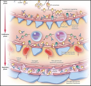
Acute Renal Failure caused by Glomerulonephritis. Acute glomerulonephritis is a type of intrarenal acute renal failure usually caused by an abnormal immune reaction that damages the glomeruli. In about 95 per cent of the patients with this disease, damage to the glomeruli occurs 1 to 3 weeks after an infection elsewhere in the body, usually caused by certain types of group A beta streptococci. The infection may have been a streptococcal sore throat, streptococcal tonsillitis, or even streptococcal infection of the skin. It is not the infection itself that damages the kidneys. Instead, over a few weeks, as antibodies develop against the streptococcal antigen, the antibodies and antigen react with each other to form an insoluble immune complex that becomes entrapped in the glomeruli, especially in the basement membrane portion of the glomeruli.
Once the immune complex has deposited in the glomeruli, many of the cells of the glomeruli begin to proliferate, but mainly the mesangial cells that lie between the endothelium and the epithelium. In addition, large numbers of white blood cells become entrapped in the glomeruli. Many of the glomeruli become blocked by this inflammatory reaction. Those that are not blocked usually become excessively permeable, allowing both protein and red blood cells to leak from the blood of the glomerular capillaries into the glomerular filtrate. In severe cases, either total or almost complete renal shutdown occurs. The acute inflammation of the glomeruli usually sub- sides in about 2 weeks, and in most patients, the kidneys return to almost normal function within the next few weeks to few months. Sometimes, however, many of the glomeruli are destroyed beyond repair; in a small percentage of patients, progressive renal deterioration continues indefinitely, leading to chronic renal failure, as described in a subsequent section of this chapter.
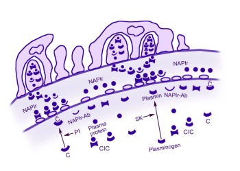
A schematic representation of the proposed mechanism for acute poststreptococcal glomerulonephritis (APSGN). C = Activated complement; Pl = Plasmin; NAPlr = Nephritis-associated plasmin receptor; SK = Streptokinase; CIC = Circulating immune complex.
Acute glomerulonephritis is a disease characterized by the sudden appearance of edema, hematuria, proteinuria, and hypertension. It is a representative disease of acute nephritic syndrome in which inflammation of the glomerulus is manifested by proliferation of cellular elements secondary to an immunologic mechanism.
Acute poststreptococcal glomerulonephritis (APSGN) results from an antecedent infection of the skin or throat caused by nephritogenic strains of group A beta-hemolytic streptococci.The concept of nephritogenic streptococci was initially advanced by Seegal and Earl in 1941, who noted that rheumatic fever and acute poststreptococcal glomerulonephritis (both nonsuppurative complications of streptococcal infections) did not simultaneously occur in the same patient and differ in geographic location.Acute poststreptococcal glomerulonephritis occurs predominantly in males and often completely heals, whereas patients with rheumatic fever often experience relapsing attacks.
The M and T proteins in the bacterial wall have been used for characterizing streptococci. Nephritogenicity is mainly restricted to certain M protein serotypes (i.e. 1, 2, 4, 12, 18, 25, 49, 55, 57, and 60) that have shown nephritogenic potential. These may cause skin or throat infections, but specific M types, such as 49, 55, 57, and 60 are most commonly associated with skin infections. However, not all strains of a nephritis-associated M protein serotype are nephritogenic.In addition, many M protein serotypes do not confer lifetime immunity. Group C streptococci have been responsible for recent epidemics of APSGN (eg, Streptococcus zooepidemicus). Thus, it is possible that nephritogenic antigens are present and possibly shared by streptococci from several groups.
In addition, nontypeable group A streptococci are frequently isolated from the skin or throat of patients with glomerulonephritis, representing presumably unclassified nephritogenic strains.The overall risk of developing acute poststreptococcal glomerulonephritis after infection by these nephritogenic strains is about 15%. The risk of nephritis may also be related to the M type and the site of infection. The risk of developing nephritis infection by M type 49 is 5% if it is present in the throat. This risk increases to 25% if infection by the same organism in the skin is present.
3. What are the different Types of the disease?
Focal Segmental Glomerulonephritis (FSGS) presents as a nephrotic syndrome. It is associated with reflux nephropathy, Alport syndrome, heroin use, or HIV. It may also be primary. The name describes its pathology—it affects only certain foci of glomeruli and then only a segment of the glomerulus. The lesions are a sclerotic glomerulus and hyalinisation of the feeding arterioles. Steroids have often been tried but have not been effective. Half of people with FSGS end up with renal failure.
Membranous glomerulonephritis presents as a nephrotic syndrome. It is the leading cause of nephrotic syndrome in adults. It is associated with cancers although most of the time it is idiopathic. In this disease, the basement membrane is thickened, but there is no significant cellularity. As GN continues, the kidney atrophies. Steroids are indicated if the disease is progressive.
IgA, Berger’s nephropathy, is the most common form of proliferative GN as well as the most common form of GN worldwide. It usually presents with hematuria. It occasionally progresses to nephrotic syndrome. One of the more common presentations is in a young male following a URI. Ace inhibitors are the primary treatment because immunosuppression is ineffective.
HSP is a variant of IgA nephropathy. Its pathology is a vasculitis of small vessels.
Post-infectious GN is the AGN associated with streptococcal infections.
Mesangiocapillary GN is associated with SLE, viral hepatitis, and hypocomplementemia.
Rapidly progressive GN, also called crescentic GN is a rapidly progressive disease that includes Goodpasture’s Syndromem, Wegener’s granulomatosis, and polyarteritis.
4. What is “acute nephritic syndrome” ?
5. What are the gross (macroscopic) findings in AGN?
6. What are the urinary abnormality seen in AGN?
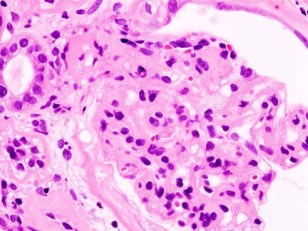 Acute nephritic syndrome is a group of disorders that cause inflammation of the internal kidney structures (specifically, the glomeruli).
Alternative Names: Glomerulonephritis – acute; Acute glomerulonephritis; Nephritis syndrome – acute
Acute nephritic syndrome is often caused by an immune response triggered by an infection or other disease.
Causes seen more frequently in children and adolescents include the following:
Associated diseases seen more frequently in adults include:
The inflammation disrupts the functioning of the glomerulus, which is the part of the kidney that controls filtering and excretion. This disruption results in blood and protein appearing in the urine, and the build up of excess fluid in the body. Swelling results when protein is lost from the blood stream. (Protein maintains fluid within the blood vessels, and when it is lost the fluid collects in the tissues of the body). Blood loss from the damaged kidney structures leads to blood in the urine.
Acute nephritic syndrome may be associated with the development of high blood pressure, inflammation of the spaces between the cells of the kidney tissue, and acute kidney failure.
Symptoms
- Blood in the urine (urine appears dark, tea colored, or cloudy)
- Blurred vision
- Decreased urine volume (little or no urine may be produced)
- General aches and pains ( joint pain, muscle aches)
- General ill feeling (malaise)
- Headache
- Slow, sluggish, lethargic movement
- Swelling of the face, eye socket, legs, arms, hands, feet, abdomen, or other areas
Late symptoms include the following:
- Signs and tests
Your blood pressure may be high. When examining your abdomen, your health care provider may find signs of fluid overload and an enlarged liver. When listening to your lungs and chest with a stethoscope, abnormal heart and lung sounds may be heard. The neck veins may be enlarged from increased pressure.
Generalized swelling is often present. There may be signs of acute kidney failure.
Tests that may be done include:
A kidney biopsy reveals inflammation of the glomeruli, which may indicate the cause.
Tests to determine the cause of the acute nephritic syndrome may include:
Treatment
The goal of treatment is to reduce the inflammation. Hospitalization is required for diagnosis and treatment of many forms of acute nephritic syndrome. The cause must be identified and treated. This may include antibiotics or other medications or treatment.
Bedrest may be recommended. The diet may include restriction of salt, fluids, and potassium. Medications to control high blood pressure may be prescribed. Corticosteroids or other anti-inflammatory medications may be used to reduce inflammation.
Other treatment of acute kidney failure may be appropriate.
Prevention
Many times the disorder cannot be prevented, although treatment of illness and infection may help to reduce the risk.
7. Why is there edema in AGN?
AGN is characterized by edema which is generalized & also known as anasarca or dropsy.
Anasarca, also known as “extreme generalized edema” is a medical condition characterised by widespread swelling of the skin due to effusion of fluid into the extracellular space.
It is usually caused by liver failure (cirrhosis of the liver) or renal failure/disease and severe malnutrition/protein deficiency. The increase in salt and water retention caused by low cardiac output can also result in anasarca as a long term maladaptive response.
8. Differentiate primary and secondary AGN syndromes.
Nephrotic syndrome has many causes and may either be the result of a disease limited to the kidney, called primary nephrotic syndrome, or a condition that affects the kidney and other parts of the body, called secondary nephrotic syndrome.
Primary
Primary causes of nephrotic syndrome are usually described by the histology, i.e. minimal change disease (MCD) like minimal change nephropathy which is the most common cause of nephrotic syndrome in children, and Focal Segmental Glomerulosclerosis which is the most common cause of nephrotic syndrome in adults.
They are considered to be “diagnoses of exclusion“, i.e. they are diagnosed only after secondary causes have been excluded.
Secondary
Secondary causes of nephrotic syndrome have the same histologic patterns as the primary causes, though may exhibit some differences suggesting a secondary cause, such as inclusion bodies.
They are usually described by the underlying cause.
Secondary causes by histologic pattern:
Membranous nephropathy (MN):
Focal segmental glomerulosclerosis (FSGS)
9. What are the clinical signs and symptoms of AGN?
Signs and symptoms of glomerulonephritis may depend on whether you have the acute or chronic form, and the cause. Your first indication that something is wrong may come from symptoms or from the results of a routine urinalysis. Signs and symptoms may include:
- Pink or cola-colored urine from red blood cells in your urine (hematuria)
- Foamy urine due to excess protein (proteinuria)
- High blood pressure (hypertension)
- Fluid retention (edema) with swelling evident in your face, hands, feet and abdomen
- Fatigue from anemia or kidney failure
10. What are the routine and serochemical tests that should be requested on this particular cases?
Specific signs and symptoms may suggest glomerulonephritis, but the condition often comes to light when a routine urinalysis is abnormal. Tests to assess your kidney function and make a diagnosis of glomerulonephritis include:
- Urine test. A urinalysis may show red blood cells and red cell casts in your urine, an indicator of possible damage to the glomeruli. Urinalysis results may also show white blood cells, a common indicator of infection or inflammation, and increased protein, which may indicate nephron damage. Other indicators, such as increased blood levels of creatinine or urea, are red flags.
- Blood tests. These can provide information about kidney damage and impairment of the glomeruli by measuring levels of waste products, such as creatinine and blood urea nitrogen.
- Imaging tests. If your doctor detects evidence of damage, he or she may recommend diagnostic studies that allow visualization of your kidneys, such as a kidney X-ray, an ultrasound examination or a computerized tomography (CT) scan.
- Kidney biopsy. This procedure involves using a special needle to extract small pieces of kidney tissue for microscopic examination to help determine the cause of the inflammation. A kidney biopsy is almost always necessary to confirm a diagnosis of glomerulonephritis.
Sources: Medical Physiology Guyton & Hall, Medscape.com, Wikipedia, Mayoclinic.com, Medhelp.org, Studentdoc.com
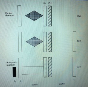 On auscultation (Figure), S1 is normal and S2 is split normally on inspiration. There is a prominent S4. A mid to late systolic ejection murmur is heard at the left sternal edge and apex. The murmur typically increases with Valsalva maneuver. When mitral regurgitation is present, an apical, holosystolic murmur is heard and may be accompanied by an S3.


