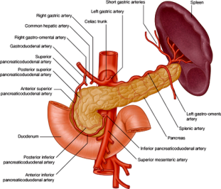
A 52-year-old Caucasian male is brought to the ER after repeated coffee-ground vomiting. After initial stabilization, endoscopy is performed revealing a deep, bleeding ulcer on the posterior wall of the duodenal bulb. This ulcer has most likely penetrated what artery/arteries?
Explanation
Duodenal ulcers are more common than gastric ulcers. Duodenal ulcers tend to occur anteriorly. Notably, ulcers located on the anterior wall of the duodenal bulb are more prone to perforation, while those on the posterior wall are more likely to cause hemorrhage.
These complications are explained by the relationship of the duodenal bulb to adjacent organs. The bulb begins at the pylorus, ends at the neck of the gallbladder, and rests anterior to the gallbladder and liver. The garstroduodenal artery, common biliary duct, and vena pora lie behind the duodenal bulb; the head of the wall, the ulcer is likely to corrode the gastroduodenal artery.
The garstroduodenal artery arises from the proper hepatic artery and perfuses both the pylorus and the proximal part of the duodenum. Damage to the gastrodudenal artery can cause significant upper gastrointestinal bleeding.
USMLE World 2009


